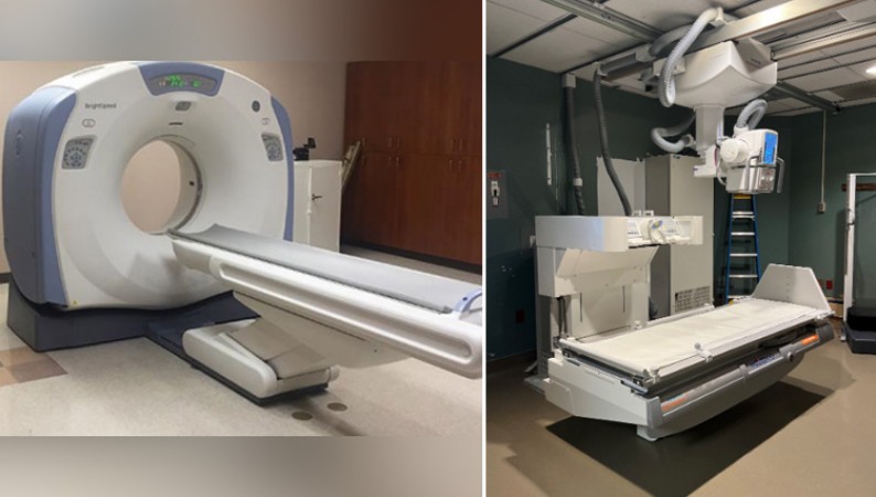
New Delhi: X-rays and CT scans are useful medical imaging tools that can offer crucial diagnostic data. However, since ionising radiation is present in both CT scans and X-rays, it is generally advised to limit unnecessary exposure. Here are a few causes for this:
1. Radiation risk: Repeated exposure to ionizing radiation from CT scans and X-rays can increase the risk of developing cancer over time. While the individual risk from a single scan is low, unnecessary or excessive imaging may lead to cumulative radiation doses that can pose a greater risk.
2.Alternative options: In some cases, alternative imaging techniques such as ultrasound or magnetic resonance imaging (MRI) can provide similar diagnostic information without using ionizing radiation. These options should be considered when appropriate.
Also Read: What makes Face Book Meta doesn't work as Expectations?
3.Radiation-sensitive populations: Certain individuals, such as pregnant women and children, are more susceptible to the potential harmful effects of radiation. Extra caution should be exercised in these cases to minimize their exposure.
4. Effective utilization: It is important to ensure that CT scans and X-rays are used judiciously, based on the clinical need and expected benefits. Unnecessary imaging can lead to increased healthcare costs, potential false positives or incidental findings, and unnecessary follow-up procedures.
While CT scans and X-rays are useful for diagnosing and monitoring a variety of conditions, it is crucial that medical professionals carefully balance any potential advantages with the radiation risks and take into account alternate imaging options whenever practical.
Also Read: Apple and Samsung looking to expand in India: R. Chandrasekhar
How much radiation a person would typically ingest from a single X-ray or CT scan?
The amount of radiation a person absorbs during an X-ray or CT scan depends on several factors, including the type of scan, the area of the body being examined, and the specific imaging protocol used. Radiation exposure is typically measured in units of millisieverts (mSv), which quantifies the absorbed dose of radiation.
For a typical single X-ray, the effective dose can range from around 0.01 mSv to 0.1 mSv, depending on the specific procedure and body part imaged. Dental X-rays, for example, usually result in a lower radiation dose compared to chest X-rays.
CT scans generally involve higher radiation doses compared to X-rays. The effective dose from a CT scan can range from a few mSv to over 20 mSv, depending on the scan type (e.g., head, abdomen, chest) and the specific imaging parameters used. Some complex CT scans, such as those with contrast agents or multiple phases, can result in higher radiation doses.
It's important to note that these radiation doses are approximate values and can vary based on the equipment and techniques used in different medical facilities. Radiology departments strive to optimize imaging protocols to minimize radiation exposure while still obtaining the necessary diagnostic information.
Also Read: App developers and start-ups have expressed their disapproval of Google's new payment policies
To put these doses into perspective, the average background radiation that a person receives from natural sources (such as cosmic rays and radon gas) is approximately 2-3 mSv per year. It's also worth noting that the risk of developing cancer due to radiation exposure increases with higher doses, but the absolute risk from a single diagnostic X-ray or CT scan is considered relatively low.
It's always recommended to discuss any concerns about radiation exposure with your healthcare provider, who can provide personalized information based on your specific situation and medical needs.