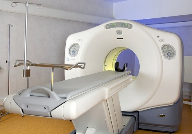
A PET-CT scan combines two imaging techniques: Positron Emission Tomography (PET) and Computed Tomography (CT). This fusion provides detailed information about the structure and function of cells and tissues in the body.
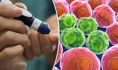
A PET scan uses a small amount of radioactive glucose to detect metabolic activity, while a CT scan provides detailed anatomical images. Together, they give a comprehensive view of the body’s internal workings.
The combination allows for more precise diagnosis and treatment planning by correlating functional and anatomical data, improving accuracy in detecting cancerous cells.
Patients are typically advised to fast for several hours before the scan. Staying hydrated is essential, and specific instructions regarding medications may be given.
The patient receives an injection of a radioactive tracer, commonly fluorodeoxyglucose (FDG). After allowing time for the tracer to distribute throughout the body, the patient lies on a table that slides into the PET-CT scanner.
The CT scan is performed first, followed by the PET scan. The entire process usually takes about 30 minutes to an hour, during which patients must remain still.
After the scan, patients are encouraged to drink plenty of fluids to help flush the radioactive material from their system. Normal activities can typically be resumed immediately.
Cancer cells metabolize glucose at higher rates than normal cells. The PET scan highlights these areas of increased metabolic activity, often indicating the presence of cancer.
PET-CT scans are instrumental in staging cancer by showing whether it has spread to other parts of the body, helping to determine the most effective treatment plan.
Doctors use PET-CT scans to monitor the effectiveness of cancer treatments, such as chemotherapy and radiation, by observing changes in metabolic activity and tumor size.
Regular PET-CT scans can detect the recurrence of cancer at an early stage, often before symptoms appear.
Combining PET and CT scans improves the accuracy of cancer detection and diagnosis by providing both metabolic and anatomical information.
PET-CT scans can detect cancer early, sometimes even before structural changes occur, allowing for prompt treatment.
These scans provide a full-body overview, useful in assessing the spread of cancer and planning treatment.
PET-CT scans are non-invasive and generally well-tolerated by patients, with minimal discomfort.
While PET-CT scans involve exposure to radiation, the levels are generally considered safe and are outweighed by the diagnostic benefits.
There is a risk of false positives (indicating cancer where there is none) and false negatives (failing to detect cancer), which necessitates follow-up testing and consultation.
PET-CT scans can be expensive and may not be available in all medical facilities, potentially limiting access for some patients.
Research is ongoing to develop new tracers that can provide more specific information about different types of cancer.
Advancements in imaging technology are continually improving the resolution and accuracy of PET-CT scans.
AI is being integrated into PET-CT scanning to enhance image analysis, providing quicker and more accurate interpretations.
Follow the instructions provided by your healthcare provider regarding fasting, hydration, and medications. Wear comfortable clothing and remove any metal objects.
Understanding the procedure and its benefits can help alleviate anxiety. Discuss any concerns with your medical team beforehand.
It is crucial to remain as still as possible during the scan to ensure clear images. The scanning table will move slowly, and the process is usually painless.
You will be able to communicate with the technician throughout the scan. Inform them immediately if you experience any discomfort.
Drink plenty of fluids to help eliminate the radioactive tracer from your body.
Your doctor will discuss the results with you and outline any necessary follow-up steps or treatments.
The detailed images from PET-CT scans allow for highly customized treatment plans tailored to the specific type and stage of cancer.
Surgeons use PET-CT scans to plan procedures with greater precision, targeting cancerous tissues while sparing healthy ones.
Radiation therapists use the detailed images to direct radiation precisely to cancerous areas, minimizing damage to surrounding healthy tissue.
Many insurance plans cover PET-CT scans, especially when deemed medically necessary. Check with your provider for specific coverage details.
Discuss potential out-of-pocket costs with your healthcare provider and explore financial assistance options if needed.
Ongoing research is expanding the use of PET-CT scans in oncology, including applications in immunotherapy and personalized medicine.
Efforts are being made to make PET-CT scans more widely available, particularly in underserved regions, to improve global cancer care.
Which Porsche car is the most powerful? Speed of 100kmph in 2.1 seconds
Sachin Khilari Defends Gold at World Para Athletics, India Achieves Best-Ever Medal Tally
This new plan of Jio will last for 365 days, you will get free FanCode along with unlimited calling